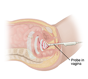
A transvaginal ultrasound is an imaging test. An ultrasound uses sound waves to form pictures of your organs that can be seen on a screen. It's often done with a probe placed on the belly. A transvaginal ultrasound uses a special probe that is put into your vagina. It uses sound waves to make pictures of your uterus, ovaries, and other pelvic organs. This test can be used to check symptoms such as pain. It can also check for problems. In pregnant women, it's used to check the unborn baby or fetus. The test is often done by a specially trained technologist called a sonographer.
Getting ready for your test
-
You may be asked to go to the bathroom. Your bladder may need to be empty before the test.
-
Tell the sonographer what medicines you take. Let them know if you have had pelvic surgery.
-
Answer any other questions the sonographer asks. Your answers will help them adapt the test to your health needs.
During your test
-
You may change into a hospital gown. You'll then lie down on an exam table with your knees raised, as you would for a pelvic exam.
-
The sonographer will use a thin handheld probe. It's shaped like a tampon. The probe has a sterile cover and nongreasy gel. It's gently put inside your vagina. In some cases, you may be asked to put the probe in yourself, as you would a tampon.
-
The sonographer moves the probe to get the best picture. You may feel pressure. Tell the sonographer if you feel pain.
After your test
Before leaving, you may need to wait for a short time while the images are reviewed. You can go back to your normal routine right after the test. Your doctor will let you know when the results of your test are ready.
Note
Be aware that although the sonographer can answer questions about the test, only your doctor can explain the results.

OB/GYN
Find a DoctorRelated Articles
Magnetic Resonance Imaging (MRI)
Magnetic resonance imaging (MRI) is a test that lets your doctor see detailed pictures of the inside of your body. MRI combines the use of strong magnets and radio waves to form an MRI image. Read on to learn all about having an MRI.
PET Scan
A PET scan can show changes in how an organ or tissue works. This can help your healthcare provider diagnose problems and create a treatment plan for you. Here's what to expect from this procedure.
Ultrasound
Ultrasound imaging is a test that uses sound waves to make detailed pictures of your organs. There are three different kinds of ultrasound imaging: abdominal, pelvic, and Doppler ultrasound. Each of these imaging tests can help your healthcare provider assess pain or other symptoms in different parts of your body. Ultrasound does not involve any radiation, and is generally a very safe procedure. Still, you should discuss any concerns with your healthcare provider.
Breast MRI
Magnetic resonance imaging creates detailed images of the body using large magnets and a computer. For breast MRI, a woman will lie face down with her breasts positioned through holes in a table.

