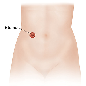
Urostomy is surgery that provides a new way for the body to get rid of pee (urine). It's done when the bladder is diseased or damaged. During the surgery, the surgeon brings part of the urinary tract or some of the digestive tract through the belly (abdominal) wall. A small opening called a stoma is made in the abdomen. This allows pee and mucus to pass out of the body. A urostomy can be done in any of the ways described below.
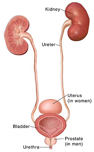
The urinary tract
Your urinary tract rids the body of liquid waste known as pee (urine). The urinary tract has many parts. It has 2 kidneys, 2 tubes called ureters, the bladder, and a tube called the urethra.
The kidneys filter waste substances and extra water from the blood. This makes pee. The 2 ureters send pee from the kidneys to the bladder. The bladder stores pee. The urethra releases pee from the bladder to outside the body.
Common types of urostomy
These include:
-
Ileal conduit. This surgery makes a tube (conduit) from part of the ileum. This is the last part of the small intestine. Pee leaves the body through this tube. One end of the conduit is sewn shut. The other end is brought through the abdominal wall to form a stoma. The ureters are detached from the bladder and connected to the conduit. Pee flows through the ureters and into the conduit. Pee then leaves the body through the stoma. This surgery doesn't change the way poop passes from the body. An ileal conduit is the most common type of urostomy.
-
Colon conduit. This surgery is done like an ileal conduit. But the tube is made from a piece of the colon and not the ileum. The stoma is bigger, as the colon is wider than the ileum.
-
Ureterostomy. This surgery brings the ureters through the abdominal wall to form 1 or 2 small stomas. The stomas are small because the ureters are much more narrow than the ileum or the colon.
-
Continent cutaneous diversion. A pouch may be made under the abdominal skin using tissue from the stomach or intestines. Pee collects in the pouch. It stays in the pouch because of a valve. You don't wear a bag. Instead, a thin tube (catheter) is used to empty the pouch when needed.
-
Orthotopic neobladder. For some people, it may be possible to make a new bladder from a part of the bowel. The new bladder is connected to the urethra. You can then pee normally. No stoma is needed. The pee empties naturally through the urethra. If the new bladder doesn’t work as a normal bladder, a catheter may have to be inserted to drain pee.
Understanding a stoma
A stoma is an opening on the abdomen through which pee and mucus can pass. It's made by bringing the end of the ileum, the colon, or 1 or both ureters through the abdominal wall. This end is then turned back on itself, like a cuff.
-
The stoma is pink or red and moist. This is because the insides of the ileum, the colon, and the ureters are like the inside of the mouth.
-
The stoma shrinks to its final size 6 to 8 weeks after surgery. Then it will be round or oval. The stoma will either be flat or it will sit ¼ inch to ½ inch above the skin.
-
With an ileal or a colon conduit, both pee and mucus pass through the stoma. After a ureterostomy, only pee comes out of the stoma.
-
Pee draining from the stoma is often collected into an attachable pouch. Pee can be drained from the pouch at your convenience or when the pouch is full.

